

The Science and Evaluation of Whitening: Understanding Skin Whitening
Skin whitening is a cosmetic process aimed at achieving a lighter, more even skin tone by reducing melanin, the pigment responsible for skin color. Whether targeting dark spots, hyperpigmentation, or uneven tone, understanding the science behind whitening and using advanced tools to evaluate results is crucial for safe and effective treatments.
The Science of Skin Layers and Skin Whitening
To understand skin whitening, it's essential to first explore the structure of the skin and how melanin is produced:
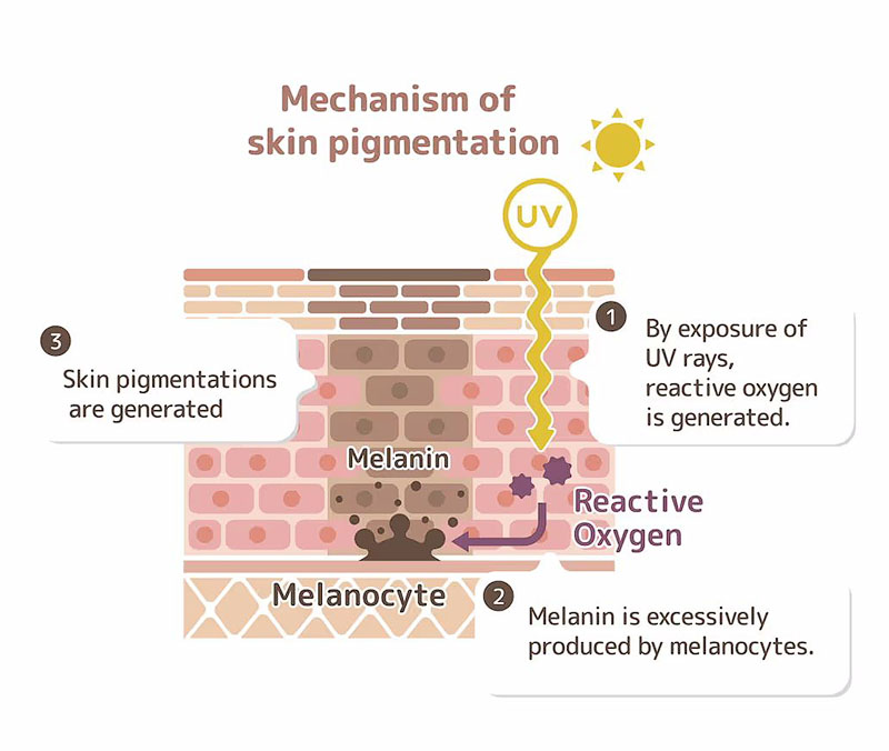
Epidermis: The outermost layer of the skin, where melanocytes (melanin-producing cells) are found. These cells produce melanin to protect deeper layers from UV radiation. Melanin accumulates in the upper layers of the epidermis, leading to visible skin tone and pigmentation. Whitening treatments primarily target this layer by inhibiting melanin production or reducing its appearance on the skin surface.
Dermis: The middle layer of the skin, which supports the epidermis but doesn't directly produce melanin. However, the health of the dermis can affect the skin’s overall appearance. Treatments that enhance dermal hydration and collagen production can help create a brighter, more even complexion by supporting the smooth, healthy surface of the epidermis.
Hypodermis: The deepest layer, composed mainly of fat and connective tissue. While not directly involved in skin whitening, maintaining overall skin health in this layer can contribute to a more youthful appearance, which enhances the results of whitening treatments.
Tools for Evaluating Skin Whitening
Accurate evaluation of skin whitening results is essential for tracking progress and ensuring safety.Several advanced tools are used to measure changes in skin pigmentation and tone.
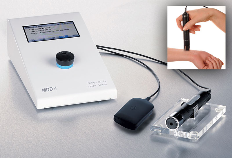
1. Mexameter: Measuring Melanin and Redness
The Mexameter is a specialized device that measures the levels of melanin and erythema (redness) in the skin.
How It Works: The Mexameter uses light of specific wavelengths to determine how much light is absorbed by melanin and hemoglobin. This provides a precise reading of pigmentation and skin redness.
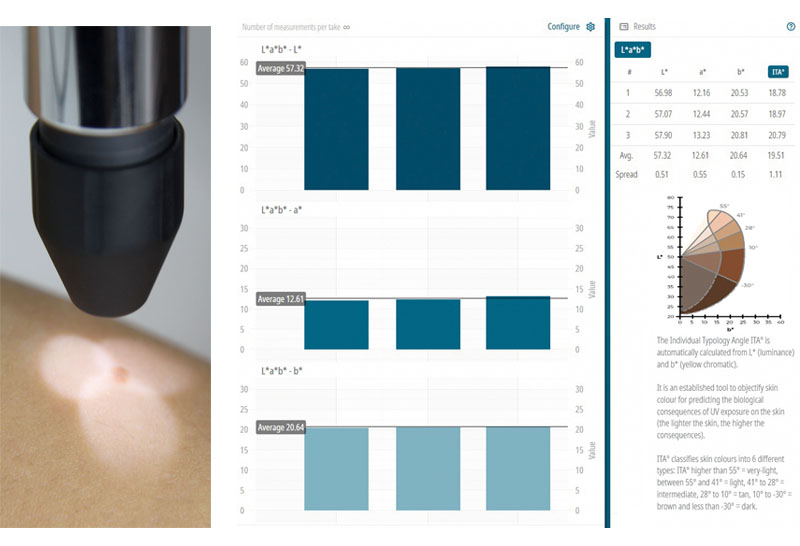
2. Colorimeter: Quantifying Skin Tone
The Colorimeter provides an objective measurement of skin color by assessing the lightness and color balance of the skin.
How It Works: It measures skin on the CIE Lab color space*, quantifying:
*L (Lightness)**: How light or dark the skin is.
a and b**: Indicating redness or yellowness in the skin.
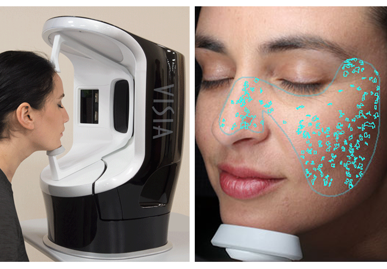
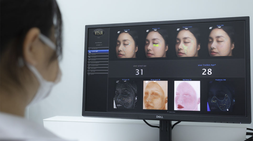
3. VISIA Imaging System: Comprehensive Skin Analysis
The VISIA Imaging System offers a full-spectrum, high-resolution analysis of the skin, capturing information on pigmentation, UV damage, and more.
How It Works: VIIS uses multi-spectral
imaging to analyze various aspects of the skin, including melanin distribution and sun damage, comparing your skin to a large database for accurate analysis.

is one of the leading research and development institute in Asia, Specializing in innovative cosmetology and oriental botanical extraction.
89 Soi Ratchataphan,
Ratchaprarop Road Makkasan,
Ratchathevee Bangkok 10400,
Thailand
© 2024 SSUP RESEARCH & INNOVATION , All Rights Reserved.